Comprehensive and Tailored Orthopedic Solutions:
Dr. Price brings a comprehensive knowledge of anatomy, deformity correction, and surgical techniques to the table. Each case of patellar luxation is carefully assessed and each individual deformity is addressed and corrected as necessary to resolve the condition. There are several common techniques employed in most cases and the most common are described below
Request a Consultation- Patellar groove assessment and deepening
– Many cases display an abnormal or shallow trochlear groove. Deepening of the groove while preserving the overlying cartilage using a wedge or block trochleoplasty will help improve patellar tracking in many cases
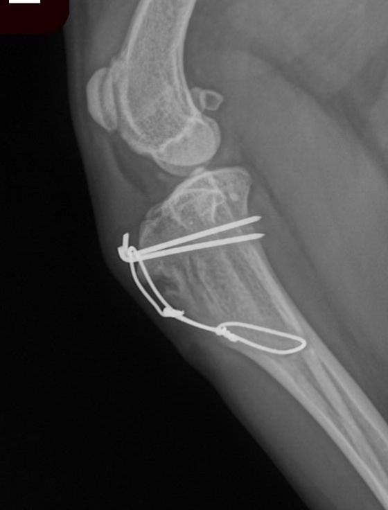
- Tibial tuberosity transposition
– The patella ligament attaches to the tibia at a point called the tibial tuberosity. This attachment point is often situated in a location medial to the centre of the joint, acting to pull the patella out of the groove. The tuberosity can be partially separated from the tibia and moved (translated) into a more lateral position. Putting the patella more in line with the groove. - Joint capsule release or imbrication
– The patella is centered within the groove by two ligaments, the medial and lateral parapatellar ligaments. Many advanced and chronic cases display secondary changes to these ligaments including excessive tightening on the medial side and laxity on the lateral side of the joint. Correcting these abnormalities further stabilizes the position of the patella within the groove. - Deformity management
– More severe cases of patella luxation display a group of deformities of the bones which render the above techniques insufficient to properly correct the disease. In these cases, repair of the deformities via distal femoral osteotomy, proximal tibial osteotomy, or combinations of these techniques can be employed. Severely abnormal patellar grooves may require a technique called patellar groove replacement. - Concurrent cruciate disease
– Grade 3/4 and 4/4 are associated with a higher risk of concurrent Cranial Cruciate Ligament (CrCL) disease. It is not uncommon to repair both patellar luxation and cruciate ligament disease at the same time. The above techniques can be combines with TPLO or other geometry modifying procedures or with extra-capsular repairs to significantly improve the comfort and quality of life of these patients. One of Dr. Price’s areas of interest involves advanced stifle surgery. He brings a high level of care and experience to these cases and commonly applies the above techniques in combination to resolve complex cases of stifle disease.